

3 min read
In vivo Acute Kidney Injury (AKI) models are commonly used to study renal damage and evaluate potential therapeutic interventions. Several models simulate AKI, including bilateral occlusion, unilateral occlusion, and nephrectomy combined with unilateral occlusion. In these models, blood is analyzed for biomarkers such as creatinine, BUN, KIM-1, and NGAL, which reflect kidney function and injury severity. Urine is tested for total protein and albumin to assess renal filtration and damage. While rodent models are commonly used, we developed a pig model that leverages the physiological similarities between pigs and humans in organ function and drug response. The comparable kidney size and function of pigs make this model highly relevant for studying renal dysfunction and testing potential therapies.
Rodent Models of AKI
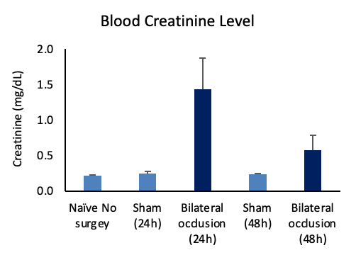
Bilateral Ischemia/Reperfusion Induced AKI in Mice

Figure 1. Blood creatinine levels in bilateral ischemia/reperfusion induced AKI in mice indicate a significant increase in blood creatinine levels following bilateral occlusion, demonstrating impaired kidney function.
Nephrectomy Followed by Unilateral Occlusion AKI in Rats
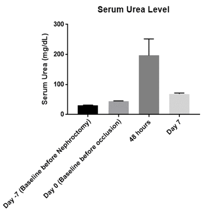
Figure 2: Serum biomarkers following nephrectomy and unilateral occlusion model shows a marked increase in serum urea post-occlusion, highlighting progressive kidney impairment.
Bilateral Occlusion-Induced AKI in Rats
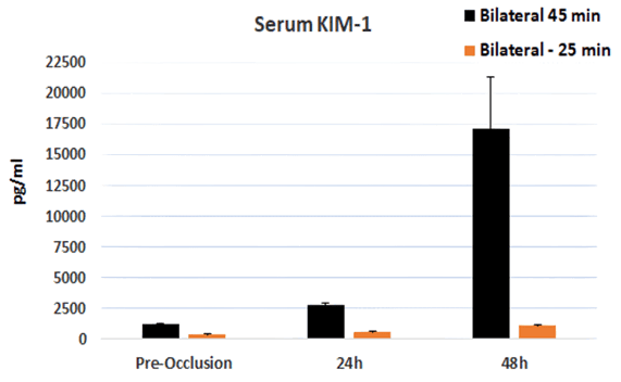
Figure 3: Serum biomarkers in bilateral occlusion in rats in 25-minute versus 45-minute occlusion show a sharp increase in KIM-1 levels, with more pronounced effects observed in the 45-minute occlusion group, indicating more severe kidney injury.
Nephrectomy and Unilateral Occlusion Induced AKI in Pigs
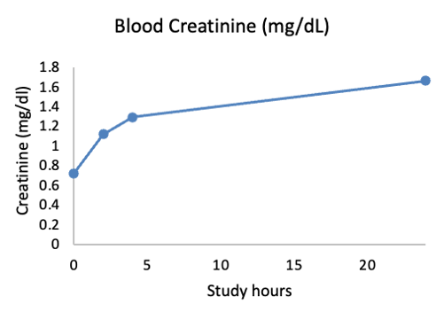
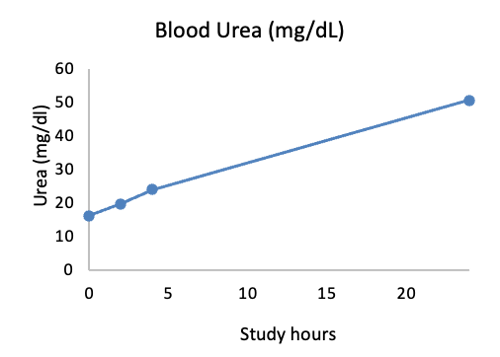
Figures 4-5: Blood creatinine and urea concentrations following AKI increase over time indicating declining kidney function post-injury.
The use of various in vivo AKI models, including both rodent and pig models, provides valuable insights into the mechanisms of kidney injury and enables the evaluation of potential therapeutic interventions. Blood biomarkers and urine analysis are essential tools for assessing renal function and injury severity. While rodent models remain widely used due to their ease of handling and low cost, the pig model offers greater relevance owing to its physiological similarities with humans.
Learn more about our acute kidney injury models by downloading our datasheet here.
Related Posts
Stress, Trauma & Pain- Image Not Imagine The Evidence| Biomarkers
The 9th Annual Congress of the European Pain Federation EFIC® was anything but painful as it...
Preclinical Human Pain Models. Translating to Clinic
Management of acute pain related to surgical intervention, termed postoperative pain, continues to...
The HTX-011 Story
HTX-011 Breakthrough in Pain Management
ZYNRELEF, the commercial name for HTX-011, is the first and...