

4 min read
Parkinson’s Disease (PD) is a progressive neurodegenerative movement disorder that affects millions of people worldwide. PD is characterized by the loss of dopaminergic neurons in the substantia nigra pars compacta (SNpc) of the midbrain. The cardinal symptoms of PD include bradykinesia, resting tremor, rigidity, and postural instability. Progress towards the identification of disease-related genes has led to the expansion of animal models of PD from the classic 1-methyl-4-phenyl-1,2,3,6-tetrahydropyridine (MPTP), 6-hydroxydopamine (6-OHDA)-induced neurotoxin models, and genetic models of the disease.
|
PD Model |
Species |
Model Length |
Assessments |
|
6OHDA PD Model |
SD Rats |
14 days |
Paw Placement, TH Immunoreactive Analysis, Biomarkers, Pharmacokinetics |
|
MPTP PD Model |
C57BL/6 Mice |
7 days |
TH Immunoreactive Analysis, Biomarkers, Pharmacokinetics |
|
Neurodegeneration Screening Assay |
Primary neurons from immature mice or rats |
5 days |
Neurodegeneration Index |
MPTP Model
- Mechanism: The MPTP model of Parkinson's disease involves the systemic administration of MPTP, a highly lipophilic compound that readily crosses the blood-brain barrier. Initially, MPTP itself is not neurotoxic but is converted to its active form, 1-methyl-4-phenylpyridinium (MPP+), within the brain. This conversion occurs via monoamine oxidase B (MAO B), leading to the preferential uptake of MPP+ by dopaminergic neurons through the dopamine transporter (DAT). Once inside dopaminergic neurons, MPP+ disrupts mitochondrial function by inhibiting complex I of the mitochondrial respiratory chain, ultimately causing cell death. The most severe impact is observed in the substantia nigra, reproducing the hallmark feature of dopaminergic neuron loss seen in PD.
- Replication of PD: This mouse model is the most commonly used PD animal model and is similar to the 6OHDA model. The MPTP model shows selective degeneration of dopaminergic neurons, closely mirroring the mechanism of PD in humans.

Figure 1. Decreased TH Immunoreactive Cells in Substantia Nigra in Acute MPTP.

Figure 2. Immunohistochemical staining for Tyrosine Hydroxylase in SNpc.
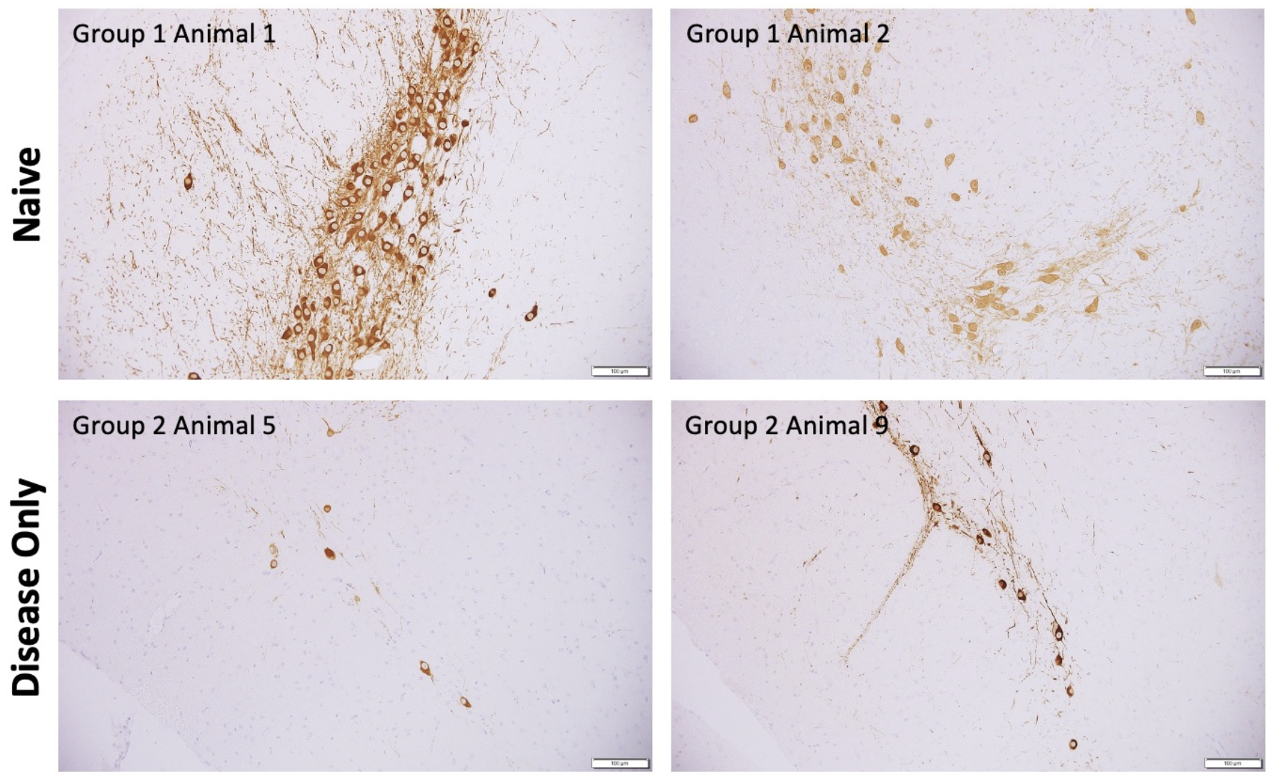
6OHDA Model
- Mechanism: The 6OHDA model of Parkinson's disease is a unilateral lesion model in which the nigrostriatal pathway is damaged. 6OHDA fails to cross the blood-brain barrier when administered systemically, so the toxin is injected stereotaxically into the brain region of interest, typically the substantia nigra (SN), ventral tegmental area (VTA) or striatum.
- Replication of PD: The 6OHDA model leads to dopaminergic neuron loss, although it's not as selective as the MPTP model in terms of targeting only dopaminergic neurons. 6OHDA injections can be targeted to specific brain regions, allowing researchers to induce lesions in different areas associated with PD symptoms, such as the striatum or substantia nigra.
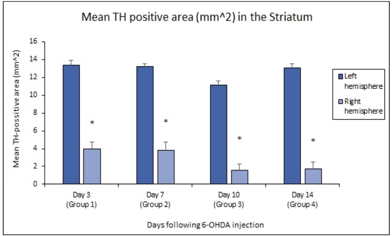
Figure 3: Changes in TH staining in the striatum due to the loss of dopaminergic neurons in the SNc.
Neurodegeneration Screening Assay
The PD in vitro screening assay utilizes primary neurons from immature mice or rats. Following the conditioning phase with alpha-synuclein, cultures are treated with compound and stained and evaluated for their neurodegeneration index. The assays are ideal for screening compounds prior to efficacy in in vivo studies.
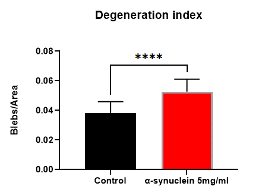
Figure 4. After 7 days of conditioning, there is an increase in appearance of blebs, indicative of neurodegeneration.
Related Posts
Promoting Diversity for Translational Success
In preclinical efficacy studies, several factors are often overlooked, and it is important to...
Increase diversity to improve clinical translation and strengthen pain management outcomes.
The goal of preclinical drug development is to identify potential therapeutic compounds and gain an...
The Critical Role of Clinical Relevance in Effective Study Design
Enhancing clinical relevance in early-stage studies can improve efficiency, reduce costs, and...