4 min read
Inflammation is a key pathological driver in many neurological diseases. The interplay between the central and peripheral nervous systems and the immune response highlights the importance of understanding the cellular and inflammatory attributes that are strongly associated with neurological diseases and pain. Biomarker analysis can strengthen CNS behavioral findings and address unanswered questions about neuroinflammation, neurodegeneration, and pain. In preclinical studies for inflammatory diseases and CNS diseases, cellular markers and cytokines can be measured in tissues, serums, or fluids and serve as critical indicators of disease progression and therapeutic response.
Biomarker assays
High throughput screening and ultra-sensitive immunoassays has made it possible to detect low levels of neuroscience and inflammatory analytes in CSF, plasma, or serum. Below are methods for detecting and evaluating neuroinflammatory markers in preclinical studies.
| Assay | What is measured |
| Flow cytometry | Cell based signatures that mirror biological aspects |
| Multiplex Analysis | Protein signatures that identify neuroinflammation and mirror other biological aspects |
| Immuno-histochemistry/ Histology | Biomarkers in tissues, intraepidermal nerve fiber (IENF) density, neuromuscular junction (NMJ) |
| In vivo Electrophysiology (EP) | Spontaneous action potentials in nerve fibers and pain-related electrical signals |
| Laser Doppler Flowmetry | Cerebral blood flow of the cortex |
Inflammatory Biomarkers
The expression of pro-inflammatory and anti-inflammatory cytokines alters the homeostasis of the central nervous system and contributes to the loss of neurons. This may lead to neurodegeneration, peripheral neuropathy, or other forms of pain. Determining which biomarkers to add to a study requires expertise on underlying inflammatory mechanisms and the resulting changes in neuronal function. Examples of common inflammatory mediators and immune cells involved in neurodegeneration and pain are listed below.
| Inflammatory Mediators | Immune cells |
|
|
Inflammatory biomarker detection in CFA-model
Acute inflammatory pain is associated with tissue injury and manifests itself as an expression of neuronal plasticity consisting of central and peripheral sensitization. Tissue injury causes a release of inflammatory mediators from damaged cells, which become central to the initiation of an inflammatory cascade. In the CFA model, it is useful to analyze pro-inflammatory and anti-inflammatory cytokines in paw skin, CSF, and plasma. Analysis of these sample types provide information on local and systemic effects of a drug.
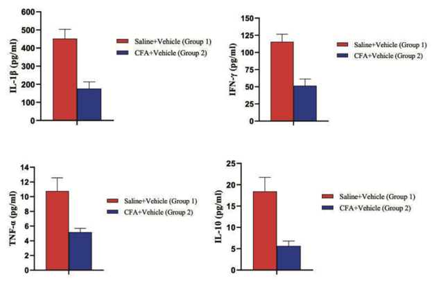
Figure 1: Pro-inflammatory and anti-inflammatory cytokines in CSF of rats. There is a reduction in TNF-a, IFN-g, IL-1b, suggesting a reduction in the pro-inflammatory response. There is also a reduction in IL-10, suggesting a reduction in the anti-inflammatory response.
Detection in STZ-induced diabetic neuropathy model
STZ is used to induce Type 1 Diabetes. Following hyperglycemia, nerve endings are damaged through an inflammatory process or interference with the blood supply.
 Figure 2: Exposure to STZ increased pro-inflammatory cytokines (TNF-α and RANTES) and reduced anti-inflammatory (IL-10) cytokines in the plasma.
Signaling cascades initiated by BDNF are key regulators of a wide range of processes in the brain, including synaptic plasticity. Changes in BDNF levels and activities have been shown to be involved in neurodegenerative conditions.
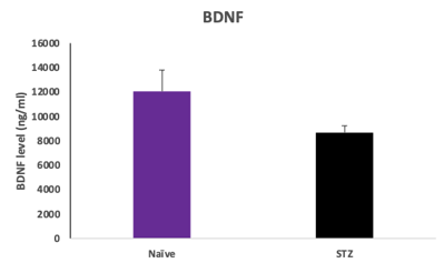
Figure 3: BDNF levels in the hippocampus 28 days following STZ. Decreased levels may show the beginning of degradation in diabetic neuropathy.
To learn more about our capabilities, contact us or download our inflammatory biomarker whitepaper below.